


Spindle Positioning in Oocytes: The Spindle View Technique
Introduction
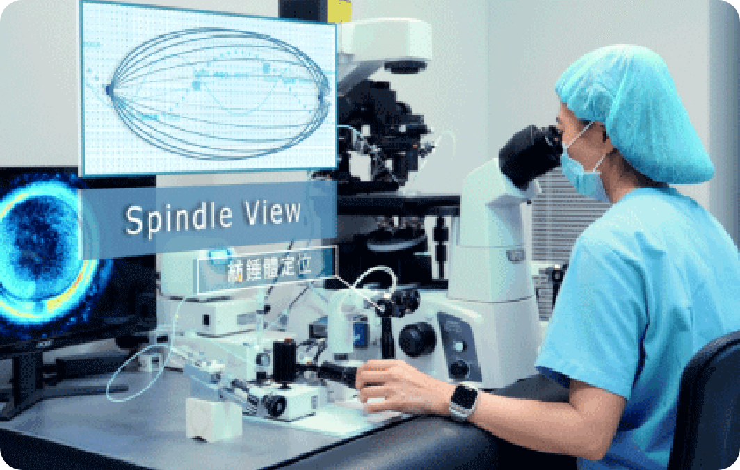
Spindle View combines polarized light microscopy with image analysis to visualize the spindle inside the oocyte in real-time, without causing harm. It enhances fertilization, improves embryo quality, and reduces chromosomal risk—especially crucial for advanced-age patients or those with diminished ovarian reserve.


Understanding the Spindle
The spindle is essential for chromosome alignment and separation during cell division. Abnormal spindle function can result in aneuploidy or genetic disorders.
Roles in Division
| Mitosis | Meiosis |
|---|---|
| Ensures daughter cells inherit the correct chromosome number. | Generates gametes (sperm or eggs) with half the chromosome set, ensuring accurate inheritance. |
Principle of Spindle Positioning in Oocytes
Polarized light microscopy leverages birefringence to visualize the spindle. During ICSI, embryologists can avoid the spindle, minimizing chromosomal damage and maximizing embryo potential.

卵子紡錘體定位對試管療程至關重要
在試管嬰兒療程(IVF)中,每一個細節都可能影響療程結果,而紡錘體定位技術正是提升受精精準度與胚胎品質的關鍵步驟。特別對於高齡、卵巢功能下降、卵子數量稀少或反覆試管失敗的患者而言,這項技術能幫助專業胚胎師避開紡錘體敏感區域,選擇最佳注射位置,增加卵子受精與發育為高品質胚胎的機會。
Expert Team
With several senior embryologists each having over 20 years of experience, Lee Women’s Hospital combines imaging interpretation with micromanipulation to ensure each oocyte is handled with the highest precision.
Who Should ConsiderThe Spindle View Technique?

Advanced maternal age women(>38 years)
Oocyte aging may lead to higher risk of spindle displacement or abnormalities. Spindle positioning can improve fertilization rates and increase the probability of generating viable embryos.

Repeated IVF failures
Couples who have undergone multiple unsuccessful IVF cycles may benefit from this technique to enhance their chances of achieving pregnancy.

Other clinical indications
In cases involving specific fertility diagnoses or compromised oocyte quality, the applicability of spindle positioning should be evaluated in consultation with a doctor.
Advanced Facilities & Techniques for Spindle Visualization
To achieve accurate observation and positioning of the meiotic spindle within oocytes, Lee Women’s Hospital employs a range of specialized instruments to ensure greater precision in laboratory procedures.
 High-resolution polarized light microscope | The fundamental tool for spindle assessment, providing clear visualization of spindle position and orientation while minimizing operator error. |
 Special glass-bottom culture dishes | Designed with high optical transparency to optimize polarized light microscopy and prevent image distortion. |
 Micromanipulation systems | Stabilize the oocyte during handling and assist in sperm injection, reducing the risk of spindle disruption. |
 Microinjectors and holding pipettes | Essential for intracytoplasmic sperm injection (ICSI), these devices provide stable pressure control, enabling precise sperm delivery into the oocyte with both safety and efficiency. |
Visual Aid
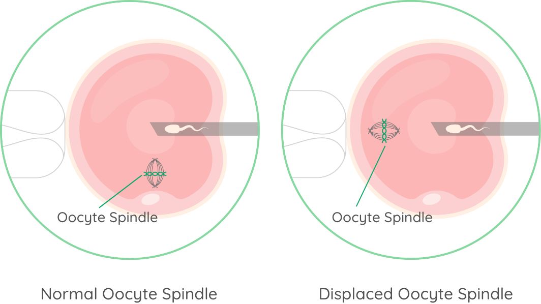
- Normal Oocyte: Predictable spindle position.
- Aged Oocyte: Spindle displacement may reduce injection accuracy without advanced technology.

Development & Hospital Advantages
2006
First adoption of spindle view technology in Taiwan
3Units
Dedicated equipment
20+ Years
of Experience Professional embryologist team
Personalized
Treatment design
The First Reproductive Center in Taiwan to Introduce Spindle Positioning Technology
Since 2006, Lee Women’s Hospital has been among the pioneers in adopting Spindle View technology, becoming one of the first medical teams in Taiwan to apply this method in clinical practice. With nearly two decades of operational experience, the hospital has accumulated extensive expertise and proven clinical outcomes.
Equipped with Advanced Polarized Light Microscopy Systems
The hospital is equipped with three dedicated spindle positioning devices for use in in vitro fertilization (IVF) procedures. These are integrated with professional image analysis systems, enabling precise observation and accurate localization of each oocyte spindle.
Highly Experienced Embryologist Team
Lee Women’s Hospital has a team of experienced embryologists, many of whom have over 20 years of hands-on experience in intracytoplasmic sperm injection (ICSI) and oocyte assessment. Their expertise ensures precise control of every detail in spindle positioning and injection procedures.
Tailored Treatment plans for Advanced-Age and Difficult Cases
Spindle View technology is particularly applied to patients of advanced maternal age, diminished ovarian reserve, or low oocyte yield. With individualized ovulation monitoring and embryo evaluation, this approach significantly enhances the likelihood of successful conception.
紡錘體定位技術,提升試管嬰兒成功率
茂盛醫院自 2006 年起,即率先引進「卵子紡錘體定位技術」應用於試管嬰兒療程,是台灣最早擁有此技術並成功導入臨床的生殖醫療機構之一。由於一套紡錘體觀測設備造價高昂,國內並非所有生殖中心皆具備此儀器與專業操作人力。目前茂盛院內配置 3 部高階偏光觀測系統,並持續投入資源,讓有需要的不孕夫妻都能有機會使用這項提升成功率的關鍵技術。
Clinical Studies
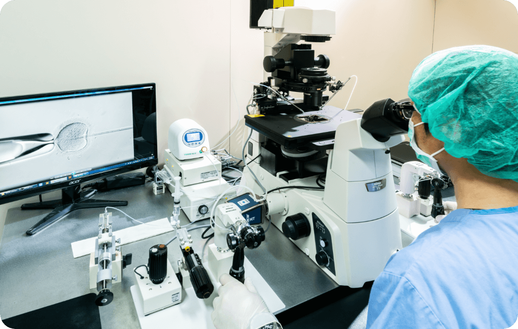
In addition to clinical practice, our team actively conducts research related to spindle positioning. In 2017, the medical team applied spindle positioning technology to patients whose intracytoplasmic sperm injection (ICSI) fertilization rates were below 50%. Results showed a significant improvement in fertilization rates. Further analysis revealed that women over 38 years of age had a markedly higher likelihood of spindle displacement away from the first polar body, a condition that may affect the accuracy of sperm injection and the developmental potential of embryos. By combining spindle imaging with ICSI, embryologists can effectively avoid cytoplasmic misalignment, thereby improving fertilization outcomes and embryo quality in advanced maternal age patients.