


AI-based Embryo Selection & Time-lapse Embryo Imaging
AI Technology for Precise Embryo Selection: Bringing Each IVF Attempt Closer to Success
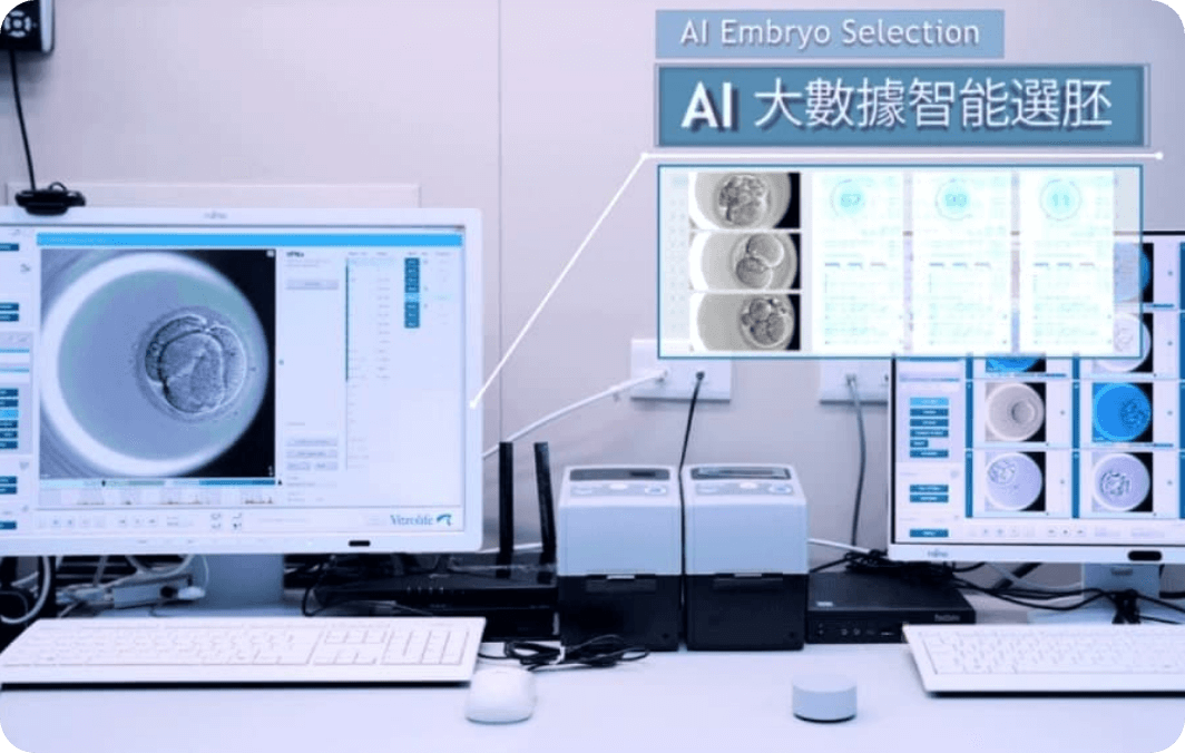
In in vitro fertilization (IVF) treatment, embryo selection is a crucial determinant of pregnancy success. Lee Women’s Hospital has pioneered the integration of the Time-lapse Imaging System with AI-based embryo selection technology. Through this approach, every detail of embryo development can be continuously monitored and analyzed with greater scientific rigor and precision.
Developing AI-based Embryo Selection: Identifying High-quality Embryos through Data

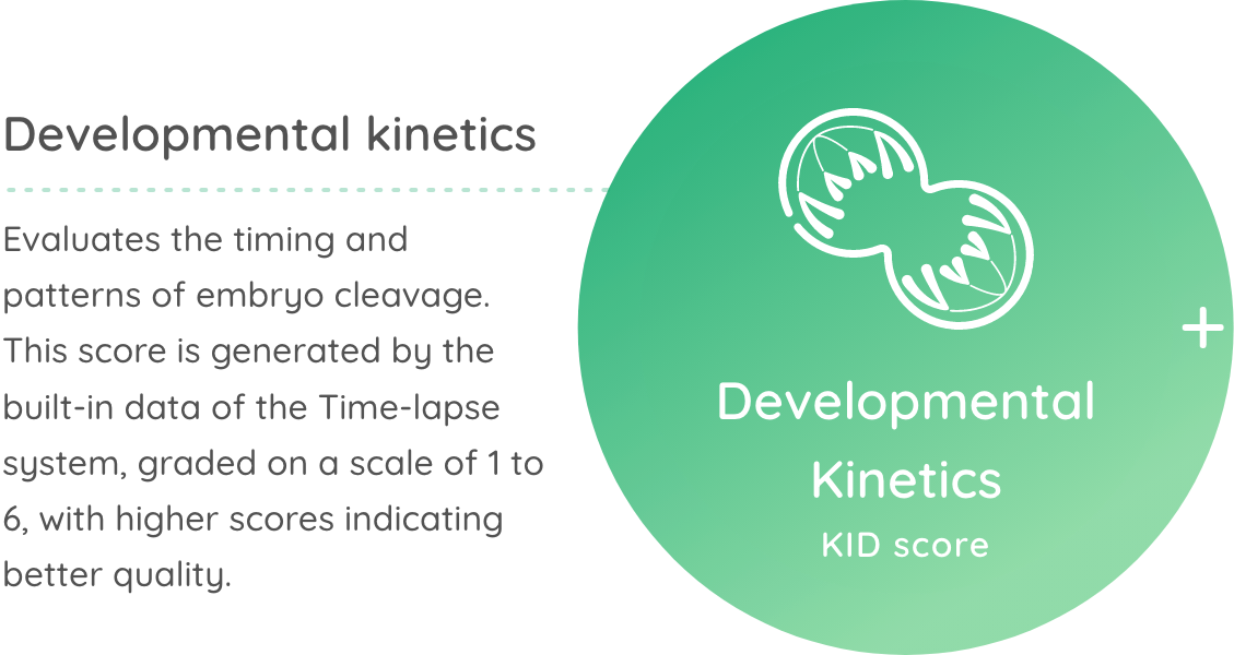


Developmental Kinetics KID score
Evaluates the timing and patterns of embryo cleavage. This score is generated by the built-in data of the Time-lapse system, graded on a scale of 1 to 6, with higher scores indicating better quality.
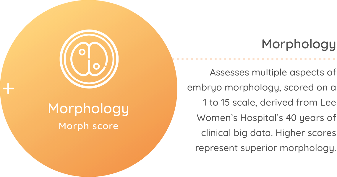
Morphology Morph score
Assesses multiple aspects of embryo morphology, scored on a 1 to 15 scale, derived from Lee Women’s Hospital’s 40 years of clinical big data. Higher scores represent superior morphology.
The combined score assists embryologists and doctors in discussing embryo grading and implantation sequence with patients.
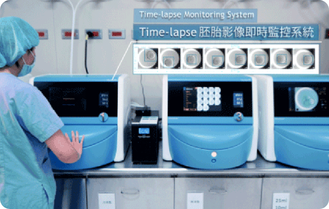
The AI system developed at Lee Women’s integrates Time-lapse embryo imaging with deep learning algorithms. By analyzing sequential images of cell divisions during embryo culture, the system predicts embryo morphology and developmental potential. This enhances embryo grading accuracy and improves the prediction of implantation outcomes, thereby increasing the overall pregnancy rate in IVF treatment.
Unlike many institutions that purchase commercial “black-box” AI systems, Lee Women’s Hospital has trained its algorithms using more than 40,000 successful IVF cases andover 4,000 annual treatment cycles. This makes the system more closely aligned with the developmental characteristics and clinical needs of Taiwanese couples with infertility, ensuring both higher precision and practical applicability.
Four Key Advantages of AI-based Embryo Selection at Lee Women’s
- Comprehensive monitoring of embryo dynamics using Time-lapse imaging combined with AI big data analysis
- Providing quantitative scoring to support doctors in formulating optimal implantation strategies
- Model trained with long-term clinical data from Lee Women’s Hospital, reflecting Asian embryo developmental patterns
- Standardized and reproducible embryo evaluation for improved clinical decision-making
Core Objectives of AI-based Embryo Selection
| Establishing an Objective and Standardized Evaluation Mechanism | Accurately Selecting Viable Embryos to Accelerate Pregnancy Goals |
|---|---|
| Through deep learning models trained on tens of thousands of embryo images, AI creates consistent and objective assessment standards. This reduces human error while improving accuracy, ensuring that every patient receives professional and unbiased embryo evaluation. | By identifying embryos with greater developmental potential, AI increases implantation success rates, shortens treatment duration, and reduces repeated embryo transfers, helping patients achieve pregnancy more efficiently. |
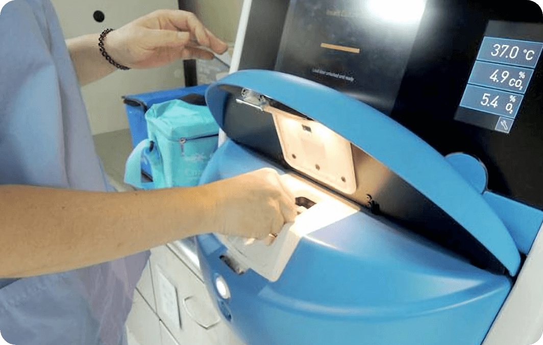
Time-lapse Embryo Imaging System
Conventional embryo observation requires opening the incubator frequently, risking changes in culture conditions. By contrast, the Time-lapse system captures embryo images every 10 minutes without disturbing the culture environment, thereby recording the entire developmental process.
These dynamic images preserve all critical events of embryo cleavage while also providing high-quality datasets for AI training, enhancing the prediction of implantation potential.
System Specifications
Imaging frequency | 11 embryo photos would be taken every 10 minutes at varying focal depths, ensuring 24-hour continuous observation. |
Resolution | Equipped with high-definition lenses to capture fine morphological details. |
Culture stability | Maintains constant temperature, humidity, pH, and gas environment without external disturbance. |
Light protection | All microscopes are fitted with green filters to reduce short-wavelength light exposure, minimizing potential embryo damage. |
Automated data analysis | AI models assess embryo development and provide quantitative scores. |
Image traceability | Complete playback of developmental stages assists embryologists in accurate evaluation. |
Advantages of Time-lapse Incubators
Conventional Incubator | Time-lapse Incubator | |
|---|---|---|
High-resolution imaging | Absent; requires opening the incubator to observe the embryos |  Present; observation without opening the incubator |
Culture environment | Stable, but disturbed during door opening | Consistently stable throughout culture period |
Monitoring | 1–2 static observations per day | Continuous 24-hour dynamic monitoring, one image every 10 minutes |
Developmental events | Many events missed | All developmental milestones recorded (up to 144 times a day) |
Embryo selection | Based on embryologist’s experience | Supported by AI algorithms combined with embryologist expertise |
Distinctions of Lee Women’s AI from Other Institutions
Unlike imported AI embryo selection tools, the Lee Women’s model is designed, trained, and validated by in-house doctors and embryologists, based on:
- Over 100 million Time-lapse embryo images combined with PGT-A outcomes for deep learning training
- Cross-validation using three embryonic developmental time partitions and three AI algorithms, generating embryo grading and implantation predictions
- Peer-reviewed publications and clinical validation, establishing Asia’s first localized AI-based embryo prediction model
Clinical Achievements
Since 2017, Lee Women’s Hospital has adopted Time-lapse embryo imaging, and in 2021 incorporated its self-developed AI selection system, establishing a clinically validated, MIT-developed big-data embryo selection model.

- The use of AI-based embryo selection with Time-lapse significantly increased pregnancy rates with single embryo transfer, especially in women over 35 years old.
- For patients with limited oocytes or unsuitable for PGT-A, AI provides a non-invasive alternative to identify embryos with higher implantation potential.
- On average, doctors reduced 1–2 transfer cycles per patient, lowering emotional stress and treatment costs.
- With over 40,000 successful IVF births accumulated, the AI model continues to improve annually through 4,000+ new treatment cycles.
This AI-embryo imaging integration has now become a standard component of IVF treatment at Lee Women’s Hospital, offering infertile couples greater confidence and opportunities for success.